At Graber & Gyllenhaal Orthodontics, we believe that technology and innovation are extremely important and have the direct ability to positively impact our patients’ experiences and outcomes at our offices. We are excited to educate you about our imaging technology which goes above and beyond the average orthodontic office.
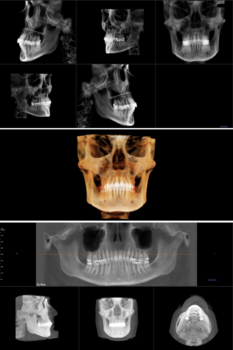
iCat® Cone Beam Computer Assembled Tomography (CBCT) imaging, also referred to as 3D radiographs or x-rays, gives us the ability to take 3D images of the teeth, jaws, bones, and facial structures at much lower energy exposure than a typical CT scan used in hospitals.

iCAT CBCT x-rays provide us the opportunity to improve diagnosis for our patients, especially in cases of impacted teeth, misdirected tooth eruption, uneven jaw growth, dental implants, and surgical orthodontics. We are able to see tooth root positions better than traditional 2D x-rays, and allows for better diagnosis and more accurate treatment planning.
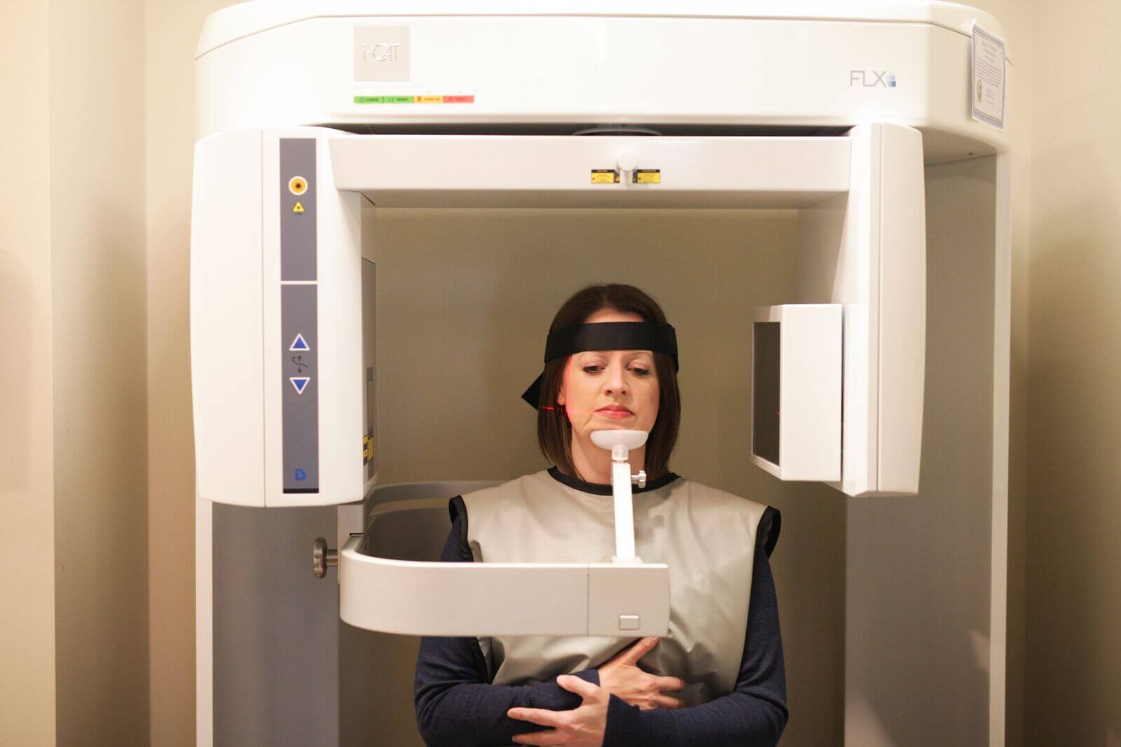
An iCat CBCT x-ray takes less than 10 seconds and only requires one image – no need for multiple x-rays and additional exposures. This single x-ray allows us to see the relationships of tooth roots, unerupted teeth, and jaw positioning from any and all angles.
This highly accurate, 3D x-ray of your teeth, jaw, and facial structures is only a fraction of energy exposure compared to traditional dental x-rays and is safe for both children and adults. Even though the energy exposure is very low, we still limit the frequency of x-rays in our office.
How much energy exposure is in one iCAT x-ray?
- 1/2 as much exposure as a full series of digital dental x-rays
- 1/5 as much as full (28) mouth series of standard dental x-rays
- 1/70 as much as a typical medical CT scan
- Less than a flight from New York to Los Angeles

Why do we take x-rays?
We take x-rays because when it comes to your overall oral health, we won’t guess. X-rays, specifically 3D x-rays, give us the best and most complete image of a patient’s current oral situation. Moving teeth without assessing the underlying tooth root position, gum health, and bone structure can be dangerous and cause irreparable damage. We work to achieve your smile goals in the safest and most comfortable way.
How are dental x-rays different than 3D x-rays?
Dental offices will often take bitewings and periodicals (PA’s), or small 2D x-rays of a small group of teeth or single tooth. Because orthodontists focus on the entire mouth and the relationship between a patient’s top and bottom jaw, we take larger x-rays that encompass the entire mouth, instead of individual teeth. iCAT CBCT images have revolutionized how we diagnose and treat orthodontic patients.
Call today to schedule your complimentary consultation to see how our 3D technology can provide more accurate treatment plans for orthodontic patients of all ages!

0 Comments